Leica SP8
This microscope is equipped with a motorized Super Z stage, motorize water cool turret, and 10X, 20X, 40X and 63X high NA objectives, capable of DIC and IR imaging. It is equipped with three laser systems for excitation: i) a white light laser (WWL2), ii) a 405 nm DMOD laser, and a iii) a 355 nm violet HeNe laser. This confocal also has the AOBS for tunable excitation for up to eight line simultaneously, all with very step transitions (<5nm). This is an extremely bright and spectrally flexible beam splitter that will allow for tremendous synergism with the white light laser. Also equipped with three HyD detectors and a resonant scanner, this confocal microscope is both extremely fast and sensitive. This confocal also has a powerful workstation with several software modules including: i) LAS X system for control of microscope hardware, ii) Live Data Mode, iii) 3D visualization tool, iv) 3D Analysis tools, v) Wizards for FRAP, FLIP, and FRET, and vi) Hyvolution for super resolution down to 140nm.
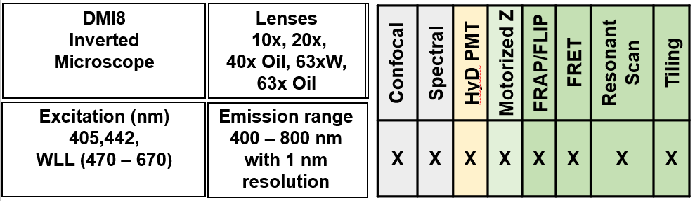
The Leica SP8 X imaging system coupled to DMI8 epi-fluorescence microscope equipped with various components as listed below-
- Objective lenses: 10X/0.40, 20X/0.70, 40X/1.25 (Oil, Ph), 63X/1.20 (Water), 63X/1.40-0.60 (Oil).
- UV laser 405 nm for imaging UV fluorophores and photoactivation.
- Laser 442 for imaging CFP, AF 430 etc.
- White light laser gives any wavelength from 470 nm to 670 nm in 1 nm increments.
- Spectral Imaging
- Six PMT detectors (5 fluorescence channels (3 conventional PMT and 2 hybrid PMTs) + 1 transmission channel).
- resonant scanner (up to 8000 Hz).
- Temperature controlled incubator.