JSM-7200 FESEM
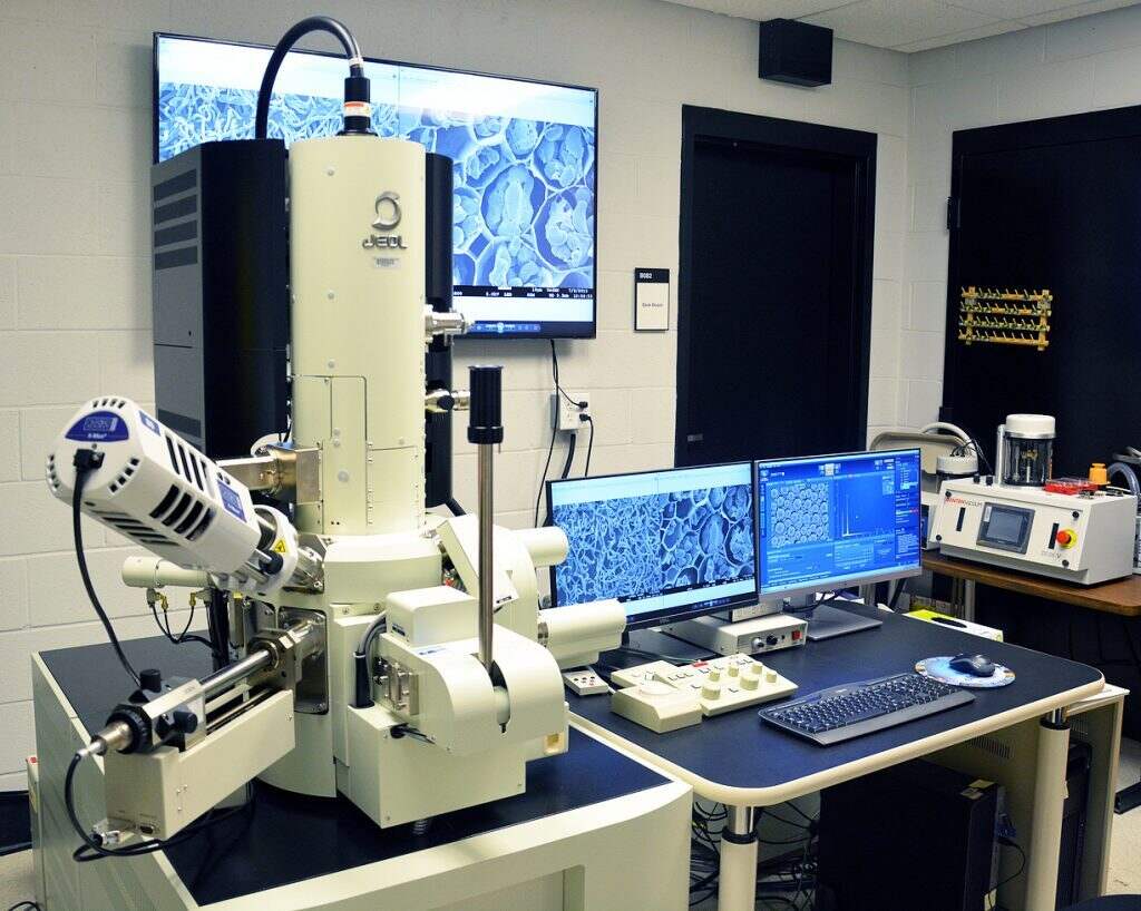
JEOL JSM-7200FLV Field-Emission Scanning Electron Microscope (FESEM)
This advanced microscope with high magnification and ultrahigh spatial resolution capabilities enables high-quality imaging and analysis of micro and nanostructures. It is equipped with multiple detectors, including an Energy Dispersive X-Ray Spectrometer (EDS), Scanning Transmission Electron Microscopy (STEM), Cathodoluminescence (CL), Back-Scattered Electron (BSE), and Secondary Electron (SE) detectors, enabling advanced analysis of various sample types.
Specifications of the FESEM:
Accelerating voltage 0.01 kV to 30 kV
Probe current 1 pA to 300 nA
Magnification Up to x1,000,000
Resolution Up to 1.2 nm
Detectors
- Secondary Electron (SE) detector
- Everhart-Thornley type in-chamber Lower electron detector (LED), and
- Through-the-lens (TTL)/Upper electron detector (UED)
- Back-Scattered Electron (BSE) detector
- Retractable BE detector (RBED) with COMPO and SHADOW modes
- Energy Dispersive X-Ray Spectroscopy (EDS) detector
- Oxford Instruments X-MaxN 80 EDS with AztecEnergy 5.0 software
- Scanning Transmission Electron Microscopy (STEM) detector
- Deben Retractable motorised Annular STEM, HAADF/MAADF/LAADF/BF, with 12 position STEM converter holder for 3mm TEM grids
- Cathodoluminescence (CL) detector
- Deben Centaurus monochromatic CL detector
Other Features
- Low vacuum (LV) capability
- Gentle Beam (GB) mode
- Charge-free (CF) scan
- Schottky-type thermal field emission gun
- 5-axis, motorized stage
- Trackball, touchpad, mouse and keyboard operation
- Stage navigation system
- Chamber scope
- 65″ LCD display
- Windows 10 Professional Operating System
- Film thickness monitor
- Data backup
- Uninterruptible power supply
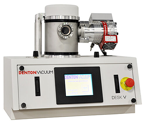
Denton Desk V TSC Sputter Coater
The Desk V sputter coater is used to prepare samples for SEM analysis. This fully automated sputter coater is designed for high-resolution electron microscopy. It is equipped with metal sputtering (Platinum) and carbon evaporation capabilities, oil-free turbo molecular pump, film thickness monitor and tilting/rotating specimen table for uniform coating. The unit is supplied with argon gas.
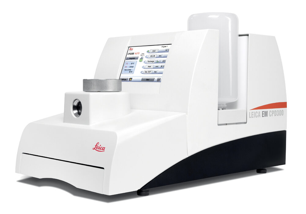
Leica EM CPD300 Critical Point Dryer
A critical point dryer is essential for preparing delicate biological specimens for SEM imaging. The Leica EM CPD300 is an advanced, fully automated unit capable of drying delicate materials for high-quality imaging. This unit uses liquid CO2(critical point 31.1 °C at 1072 psi) as a transitional medium.
Rates
User Fee
|
Services |
UM Rate |
External Academic Rate |
Non-Academic Rate |
| Sample preparation, SEM imaging and EDS analysis |
$75/hr |
$110/hr |
$150/hr (Minimum 2 hrs) |
| Hands-on training on SEM operation and sample preparation (min. 4 hrs) |
$75/hr |
$110/hr |
On request |
| Demonstration for student groups (4-5 students per group) |
$75/hr |
$110/hr |
On request |
General Policy
Before you use any instrument in the facility for the first time, you need to have a consulting tour and discussion with the core staff.
New users must request/submit an account form for approval, and approved users will have a user account.
As one of the core facilities’ aims is to encourage learning instruments and self-use, training is highly recommended. If the Core Manager assists in the experimental design, data acquisition, and scientific interpretation, they should be recognized as a co-author in the research outcomes.
Users will be charged $75/per hour for internal users (UoM) and $110/per hour for external academic users.
The rate for non-academic users is $150/per hour with a minimum of two hours. This includes assistance from lab personnel setting up and operating the SEM.
The costs of essential consumables (aluminum stubs, carbon tapes, etc.) are included in the fee. The users are responsible for any other special materials required for preparing their samples.
Booking Policy
Reservation of the instruments before use is required. The billing is based on the length of occupation time, which is figured out from the reservation and the actual log-in and out time. Please record the log-in and log-out times in the reservation system. It also helps to track the usage of instrument components.
During reserved time, the lab is unavailable for others. The reserved person can use the lab to set up the room and protocol, sample preparation, image analysis, and clean up. Users are encouraged to finalize their work within the time reserved and leave the lab promptly so that the next user can use the lab immediately.
Please let us know in advance if you are unable to use the SEM at the reserved time so others can use it.
Invoicing and Payment Policy
Invoices for services provided in the previous month will be emailed to the investigator’s fiscal contact during the second week of the month.
Payments are due within 30 days of the invoice date. If payment is not received within three billing cycles, the user cannot make new reservations until all the pending invoices are paid in full.
We strive to determine the most efficient approach to achieving each user’s research goals and will make our best effort to generate high-quality, reliable results. However, we cannot guarantee that we can produce particular results in any research project. Payment is expected for services rendered, not contingent on a positive experimental outcome.


